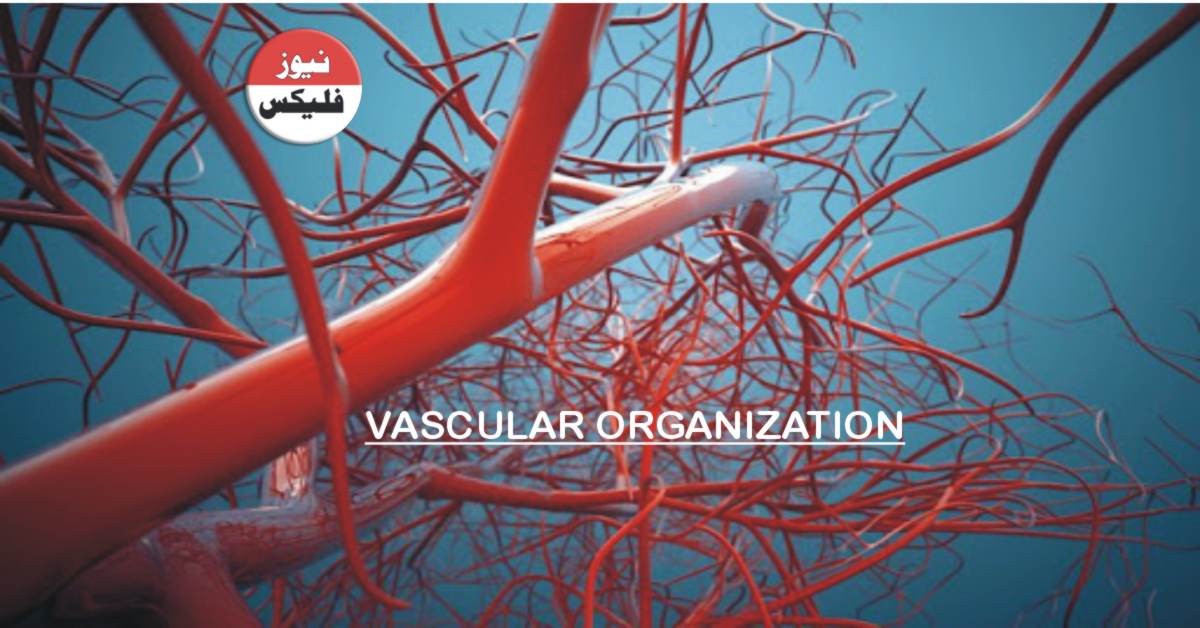
Vascular organization Arteries are divided into three types based on their size and structure:
1: Large Elastic Arteries
In these vessels are elastic fibre alternate with smooth muscle cells. throughout the media which expands during system and requires during the systole to proper blood distally. with age the elasticity is lost and vessels become stiff pipes that transmit High arterial pressures to distal organs or dilate and tortuous conduits prone to rupture.
2: Medium Sized Muscular Arteries
The media is compose primarily of smooth muscle cells with elastin limit to the internal and external elastic lamina. The medial smooth muscle cells are circularly or spirally arrange around the lumen and regional blood flow is regulate by smooth muscle cell. Contraction and relaxation controlled by the automatic nervous system and local metabolic factors.
3: Small Arteries
Small arteries live within the connective tissue of organs. The media in these vessels is mostly compose of smooth muscle cells arterioles are where blood flow resistance is regulate. As pressure drop during passage through artery walls the velocity of blood flow is sharply reduce and flow becomes steady. Together than pulsatile because the resistance to fluid flow is inversely proportional to the fourth power of the diameter small changes in arteriolar lumen size have profound effects on blood pressure.
Capillaries have lumen diameters that approximate those of red cells these vessels are line by endothelial cells and partially surround by smooth muscle cell like cells call parasites collectively capillary beds have a very large total cross sectional area and a low rate of blood flow with their thin walls and floor floor capillaries are ideally suit to the Rapid exchange of diffusible substances between blood and tissues the capillary network of most issues is necessarily very rich because diffusion of Oxygen and nutrients is not efficient beyond hundred metabolically active tissues have the highest capillary density.
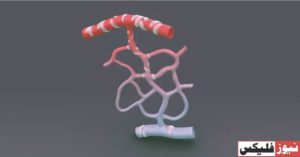
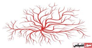
Veins receive blood from the capillary beds as postcapillary when use which anastomosis to form collecting venues and progressively larger and veins the vascular leakage and leukocyte emigration characteristic of inflammation occurs preferentially in postcapillary venublood compare with our trees at the same level of branching veins have larger diameters large Alumina and thinner walls with less distinct layers all adaptations to the low pressure is found on the side of the circulation was veins are more prone to dilation external compression and penetration by tumors or inflammatory processes in veins in which blood flows against gravity backflow is prevent by walls collectively the venous system has a huge capacitance and normally contains approximately two-thirds of the blood.
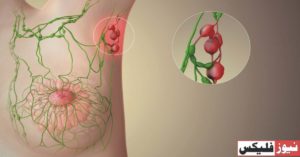
Lymphatics are thin wall endothelium line channels. That drain fluid from the interest of tissues eventually returning it to the blood. While the thoracic duct lymph also contains mono nuclear inflammatory cells and a host of proteins by delivering interstitial fluid. To lympha lymphatics enables continuous monitoring of peripheral tissues for infection. These channels can also this disseminate. This is by transporting microbes and tumor cells to distant sites.
